MDPI - Publisher of Open Access Journals
 
 
Sign in to use this feature.

Years

Between: -

Article Types

Countries / Regions

Search Results (74)

Search Parameters:
Journal = JoR

Order results
Result details
Results per page
Select all
Export citation of selected articles as:
6 pages, 698 KiB  
Case Report
Trastuzumab-Induced Organising Pneumonia in Breast Cancer Patient
by Sapna Dixit, Jyoti Bajpai, Surya Kant, Ajay Kumar Verma and Puneet Prakash
J. Respir. 2024, 4(3), 152-157; https://doi.org/10.3390/jor4030013 - 2 Jul 2024
Viewed by 283
Abstract
Patients with metastatic breast cancer often have respiratory symptoms due to various causes. Trastuzumab is a drug used in the treatment of HER2/neu over-expressing breast cancer patients. Organising pneumonia is a type of idiopathic interstitial pneumonia that mimics infection or progressive disease and [...] Read more.
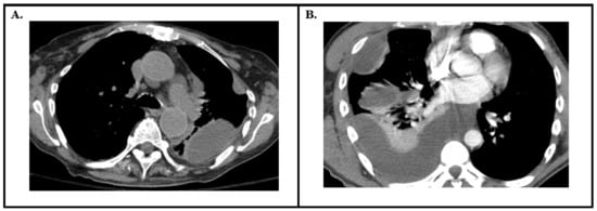
Patients with metastatic breast cancer often have respiratory symptoms due to various causes. Trastuzumab is a drug used in the treatment of HER2/neu over-expressing breast cancer patients. Organising pneumonia is a type of idiopathic interstitial pneumonia that mimics infection or progressive disease and can be difficult to diagnose in the setting of malignancy. Making a correct diagnosis is of extreme importance since any delay in the treatment can result in significant adverse patient outcome. Here, we have described a case of organising pneumonia associated with the use of trastuzumab in metastatic breast cancer patients. On the basis of clinical data, including findings such as a decreased PaO2 level and findings on chest CT scan, these patients were diagnosed with drug-induced organising pneumonia. Although it is a rare adverse event associated with trastuzumab, it may cause rapid deterioration without preceding symptoms; hence, even though it is very rare, with an incidence of less than 2%, it is still crucial to intervene so as to prevent the occurrence of such an unfavourable outcome by means of close observation and early diagnosis along with an early withdrawal of the drug and an immediate commencement of corticosteroid therapy. Full article
Show Figures

Figure 1

12 pages, 1936 KiB  
Article
Embryonic Mice with Lung-Specific RAGE Upregulation Have Enhanced Mitochondrial Respiration
by Derek M. Clarke, Katrina L. Curtis, Kaden Harward, Jared Scott, Brendan M. Stapley, Madison N. Kirkham, Evan T. Clark, Peter Robertson, Elliot Chambers, Cali E. Warren, Benjamin T. Bikman, Juan A. Arroyo and Paul R. Reynolds
J. Respir. 2024, 4(2), 140-151; https://doi.org/10.3390/jor4020012 - 5 Jun 2024
Viewed by 572
Abstract
RAGE (receptor for advanced glycation end-products) represents a class of multi-ligand pattern recognition receptors highly expressed in the vertebrate lung. Our previous work demonstrated unique patterns of RAGE expression in the developing murine lung and regulation by key transcription factors including NKX2.1 and [...] Read more.
RAGE (receptor for advanced glycation end-products) represents a class of multi-ligand pattern recognition receptors highly expressed in the vertebrate lung. Our previous work demonstrated unique patterns of RAGE expression in the developing murine lung and regulation by key transcription factors including NKX2.1 and FoxA2. The current investigation employed conditional lung-specific upregulation via a TetOn transgenic mouse model (RAGE TG) and nontransgenic controls. RAGE expression was induced in RAGE TG mice throughout gestation (embryonic day, E0-E18.5) or from E15.5-E18.5 and compared to age-matched controls. High-resolution respirometry was used to assess mitochondrial respiration and context was provided by quantifying ATP and reactive oxygen species (ROS) generation. Lung lysates were also screened by immunoblotting for MAPK/PI3K signaling intermediates. RAGE upregulation increased mitochondrial oxygen consumption in the E0-E18.5 and E15.5-E18.5 groups compared to controls. RAGE TG mice also had increased ATP concentrations, which persisted even after controlling for oxygen consumption. In contrast, ROS generation was diminished in RAGE TG animals compared to controls. Lastly, in both RAGE TG groups, pERK and pp38 were significantly decreased, whereas pAKT was significantly elevated, suggesting that RAGE signaling is likely perpetuated via pAKT pathways. Together, these data demonstrate that despite lung hypoplasia in RAGE TG mice, the remaining tissue experiences a favorable shift in mitochondrial bioenergetics without excessive redox assault and a preference for AKT signaling over ERK or p38. Full article
Show Figures

Figure 1

---

Figure 3

---

Figure 4

---

Figure 5

n = 8 mice per group). Mice were sacrificed on day E18.5 and mitochondrial respiration (A), respiratory P factor (B), and Complex II activity (C) were evaluated. Permeabilized lung tissue samples were sequentially treated with glutamate (10 mM) and malate (GM; 2 mM); +ADP (2.5 mM); +succinate (S; 10 mM); + FCCP (2 mM). Mann–Whitney tests were used, resulting in differences noted as * p ≤ 0.05 or ** p ≤ 0.01.

Full article ">p ≤ 0.05 or ** p ≤ 0.01.

Full article ">n = 4 per group). Representative blots for pAKT and actin are shown and Mann–Whitney tests revealed differences noted as * p ≤ 0.05.

Full article ">n = 4 per group). Representative blots for pERK and actin are shown and Mann–Whitney tests revealed differences noted as ** p ≤ 0.01.

Full article ">n = 4 per group). Representative blots for pp38 and actin are shown and Mann–Whitney tests revealed differences noted as ** p ≤ 0.01.

Full article ">
12 pages, 853 KiB  
Review
Robotic Bronchoscopy: A Comprehensive Review
by Bharat Singh Bhandari, Akshita Jain, Soumit Sharma, Gunjan Rana and Bruce Fernando Sabath
J. Respir. 2024, 4(2), 128-139; https://doi.org/10.3390/jor4020011 - 30 May 2024
Viewed by 350
Abstract
Lung cancer, a major global cause of cancer-related deaths, demands continual advancements in diagnostic methodologies. This review delves into the transformative role of Robotic-Assisted Bronchoscopy (RAB) in redefining lung cancer diagnostics. As lung cancer screenings intensify, leading to a surge in pulmonary nodule [...] Read more.
Lung cancer, a major global cause of cancer-related deaths, demands continual advancements in diagnostic methodologies. This review delves into the transformative role of Robotic-Assisted Bronchoscopy (RAB) in redefining lung cancer diagnostics. As lung cancer screenings intensify, leading to a surge in pulmonary nodule diagnoses, navigational bronchoscopy, notably electromagnetic navigational bronchoscopy (ENB), faces persistent limitations. Examining key RAB platforms—Monarch™, Ion™ and the Galaxy System™—reveals their distinctive features, with RAB demonstrating superior diagnostic yields over traditional biopsy methods. However, challenges include CT-to-body divergence (CBCT) and divergent findings in diagnostic yield studies and a lack of head-to-head comparisons with non-RAB modalities. Future directions should explore RAB’s potential therapeutic applications, shaping the landscape of both diagnostics and therapeutics in lung cancer management. Full article
Show Figures

Figure 1

---

Figure 3

16 pages, 827 KiB  
Review
Management of Pleural Infection: A Historical Review and Updates
by Thomas Presti, Aleezay Asghar and Nakul Ravikumar
J. Respir. 2024, 4(2), 112-127; https://doi.org/10.3390/jor4020010 - 26 Apr 2024
Viewed by 721
Abstract
Pleural infection, including empyema, continues to have a high morbidity. A deep understanding of the pathobiology and appropriate medical management is crucial to avoid complications and progression to the need for surgery. Over the last several decades, we have learned much about the [...] Read more.
Pleural infection, including empyema, continues to have a high morbidity. A deep understanding of the pathobiology and appropriate medical management is crucial to avoid complications and progression to the need for surgery. Over the last several decades, we have learned much about the pathophysiology, microbiology, and epidemiology of pleural infections. Management has changed considerably over the years with more recent clinical practices favoring minimally invasive interventions over surgery. Here we discuss in detail the pathophysiology of parapneumonic effusions as they progress from uncomplicated parapneumonic effusions to empyema and how this relates to their diagnosis and management. We review the microbiology and how it relates to recommended empiric antibiotic regimens. As intrapleural fibrinolytic therapy has become the cornerstone of management, we outline the literature on this topic dating back decades up to the most recent clinical trials and give our recommendations for management based on the literature. Full article
Show Figures

Figure 1

10 pages, 223 KiB  
Review
ICD in Cardiac Sarcoidosis: Variables Associated with Appropriate Therapy, Inappropriate Therapy, and Device Complications
by Sebastian Mactaggart and Raheel Ahmed
J. Respir. 2024, 4(2), 102-111; https://doi.org/10.3390/jor4020009 - 13 Apr 2024
Cited by 2 | Viewed by 640
Abstract
Introduction: Those with cardiac sarcoidosis (CS) are at risk of sudden cardiac death (SCD), which may be prevented using an implantable cardioverter–defibrillator (ICD). There are limited data available that follow the post-procedural outcomes of patients with cardiac sarcoidosis (CS) who have had an [...] Read more.
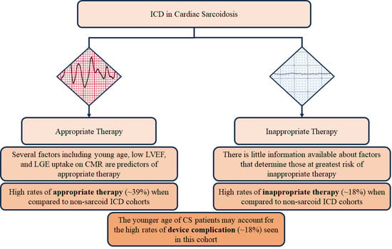
Introduction: Those with cardiac sarcoidosis (CS) are at risk of sudden cardiac death (SCD), which may be prevented using an implantable cardioverter–defibrillator (ICD). There are limited data available that follow the post-procedural outcomes of patients with cardiac sarcoidosis (CS) who have had an ICD implanted. Areas Covered: This review highlights studies that focused on both appropriate and inappropriate therapies in those with an ICD, as well as device complications in this group. There were several variables, including age, sex, ventricular characteristics, and findings on cardiac imaging that were investigated and discussed as influencing factors in predicting appropriate and inappropriate therapies. Conclusions: Adverse events in those with an ICD and CS have been minimally reported in the literature. Individuals diagnosed with CS are at high risk of ventricular arrhythmia, with comparable rates of appropriate therapy but with a higher incidence of side effects and inappropriate therapy. The younger average age of CS patients in comparison to other ICD cohorts warrants the need for further, large-scale, prospective trials with periodic interim follow-ups focused on those with this condition. Full article
Show Figures

Graphical abstract

11 pages, 536 KiB  
Article
Effect of Oscillation and Pulmonary Expansion Therapy on Pulmonary Outcomes after Cardiac Surgery
by Christopher D. Williams, Kirsten M. Holbrook, Aryan Shiari, Ali A. Zaied, Hussam Z. Al-Sharif, Abdul R. Rishi, Ryan D. Frank, Adel S. Zurob and Muhammad A. Rishi
J. Respir. 2024, 4(2), 91-101; https://doi.org/10.3390/jor4020008 - 2 Apr 2024
Viewed by 673
Abstract
Background: Oscillation and pulmonary expansion (OPE) therapy can decrease postoperative pulmonary complications in a general surgical population, but its effect after cardiac surgery has not been reported, to our knowledge. We hypothesized that using an OPE device after cardiac surgery before extubation would [...] Read more.
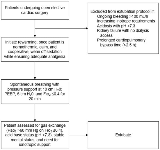
Background: Oscillation and pulmonary expansion (OPE) therapy can decrease postoperative pulmonary complications in a general surgical population, but its effect after cardiac surgery has not been reported, to our knowledge. We hypothesized that using an OPE device after cardiac surgery before extubation would decrease pulmonary complications. Methods: This retrospective cohort study included adults undergoing elective open cardiac surgery at our institution from January 2018 through January 2019, who had an American Society of Anesthesiologists score of 3 or greater. For mechanically ventilated patients after cardiac surgery, a new OPE protocol was adopted, comprising an initial 10-min OPE treatment administered in-line with the ventilator circuit, then continued treatments for 48 h after extubation. The primary outcome measure was the occurrence of severe postoperative respiratory complications, including the need for antibiotics, increased use of supplemental oxygen, and prolonged hospital length of stay (LOS). Demographic, clinical, and outcome data were compared between patients receiving usual care (involving post-extubation hyperinflation) and those treated under the new OPE protocol. The primary outcome measure was the occurrence of severe postoperative respiratory complications, including the need for antibiotics, increased use of supplemental oxygen, and prolonged hospital length of stay (LOS). Demographic, clinical, and outcome data were compared between patients receiving usual care (involving post-extubation hyperinflation) and those treated under the new OPE protocol. Results: Of 104 patients, 54 patients received usual care, and 50 received OPE. Usual-care recipients had more men (74% vs. 62%; p = 0.19) and were older (median, 70 vs. 67 years; p = 0.009) than OPE recipients. The OPE group had a significantly shorter hospital LOS than the usual-care group (mean, 6.2 vs. 7.4 days; p = 0.04). Other measures improved with OPE but did not reach significance: shorter ventilator duration (mean, 0.6 vs. 1.1 days with usual care; p = 0.06) and shorter LOS in the intensive care unit (mean, 2.7 vs. 3.4 days; p = 0.06). On multivariate analysis, intensive care unit LOS was significantly shorter for the OPE group (mean difference, −0.85 days; 95% CI, −1.65 to −0.06; p = 0.04). The OPE group had a lower percentage of postoperative complications (10% vs. 20%). Conclusions: OPE therapy after cardiac surgery is associated with decreased intensive care unit (ICU) and hospital LOS. Full article
Show Figures

Figure 1

13 pages, 539 KiB  
Review
Education in Interventional Pulmonology: How We Got Here and Where to Go from Here
by Dylan Harrell and David M. Chambers
J. Respir. 2024, 4(1), 79-90; https://doi.org/10.3390/jor4010007 - 8 Mar 2024
Viewed by 960
Abstract
Interventional pulmonology is a rapidly growing field with increasing demand. To meet this demand, training in interventional pulmonology is expanding. What started as a single training program without a standardized curriculum has grown to 40 accredited training programs with a well-defined curriculum that [...] Read more.
Interventional pulmonology is a rapidly growing field with increasing demand. To meet this demand, training in interventional pulmonology is expanding. What started as a single training program without a standardized curriculum has grown to 40 accredited training programs with a well-defined curriculum that is now overseen by the Accreditation Council for Graduate Medical Education. As the field develops, research is being actively performed to develop validated competency assessment tools and describe the learning curves for pulmonary procedures. As research evolves, this information can be used to better standardize training in interventional pulmonology and move the field towards a competency-based training model. Full article
Show Figures

Figure 1

17 pages, 2032 KiB  
Review
Airway Stents in Interventional Pulmonology
by Sami I. Bashour and Donald R. Lazarus
J. Respir. 2024, 4(1), 62-78; https://doi.org/10.3390/jor4010006 - 1 Mar 2024
Viewed by 1231
Abstract
Airway stents, first developed in the 1980s, have become fundamental in managing a multitude of airway pathologies and complications within the field of interventional pulmonology. The primary function of an airway stent is to re-establish airway patency and integrity when obstruction, stenosis, anastomotic [...] Read more.
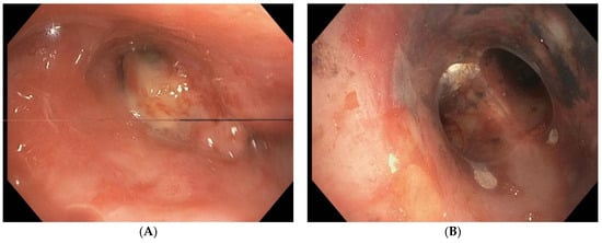
Airway stents, first developed in the 1980s, have become fundamental in managing a multitude of airway pathologies and complications within the field of interventional pulmonology. The primary function of an airway stent is to re-establish airway patency and integrity when obstruction, stenosis, anastomotic dehiscence, or fistulae develop as a result of various malignant or benign conditions. Nevertheless, airway stents are foreign bodies that can result in complications. In this review article, we will discuss airway stents and their ongoing role in the management of several malignant and benign diseases. We will describe indications for airway stenting and review the elements that must be taken into consideration for optimal patient and stent selection. Given the prevalence of data regarding therapeutic bronchoscopy and airway stenting in malignant airway obstruction, much of the discussion in this review will focus on stent placement for that indication. We will also review the data as it pertains to safety, efficacy, and complications after stent placement, and conclude with a discussion of the future applications and research avenues related to airway stents. Full article
Show Figures

Figure 1

---

Figure 3

---

Figure 3 Cont.

---

Figure 4

12 pages, 2343 KiB  
Review
Lung Cancer Staging—A Clinical Practice Review
by Ali B. Rueschhoff, Andrew W. Moore and Maykol R. Postigo Jasahui
J. Respir. 2024, 4(1), 50-61; https://doi.org/10.3390/jor4010005 - 28 Feb 2024
Viewed by 1342
Abstract
Lung cancer is the leading cause of cancer-associated death globally. Staging provides classification of the anatomic extent of cancer that is used consistently worldwide. Lung cancer staging is necessary for prognostication, to inform treatment options, and to allow accurate representation in clinical trials. [...] Read more.
Lung cancer is the leading cause of cancer-associated death globally. Staging provides classification of the anatomic extent of cancer that is used consistently worldwide. Lung cancer staging is necessary for prognostication, to inform treatment options, and to allow accurate representation in clinical trials. Staging also separates operable from inoperable disease. Since its introduction in the 1970s, the Tumor, Node and Metastasis (TNM) Staging System has undergone significant revisions, with the latest version, the eighth edition, being effective internationally since 2017. Advances in bronchoscopic and thoracoscopic technologies have expanded procedures to diagnose lung cancer and accurately define the anatomic stage. Understanding the advantages and disadvantages of available methods for staging lung cancer is critical to clinician decision making. In patients with lung cancer without distant metastases, the staging of mediastinal lymph nodes determines treatment options. To minimize the risk and cost, the most appropriate method of staging should identify the highest disease stage while carrying acceptable risk. Minimally invasive endoscopic needle techniques to stage the mediastinum are the first choice to assess for metastases in accessible lymph node stations. Surgical techniques are generally reserved for specific clinical situations, including following negative endoscopic needle techniques when suspicion for nodal involvement is high and to assess endoscopically inaccessible lymph nodes. This review provides a concise account of TNM staging of non-small cell lung cancer (NSCLC) and overview of procedures available for the staging of lung cancer. Full article
Show Figures

Figure 1

---

Figure 3

15 pages, 4594 KiB  
Review
A Review of Medical Thoracoscopy and Its Role in Management of Malignant Pleural Effusion
by Michael Gioia and Rosa L. Arancibia
J. Respir. 2024, 4(1), 35-49; https://doi.org/10.3390/jor4010004 - 26 Feb 2024
Viewed by 1303
Abstract
Pleural effusion is the most common disease among all pleural diseases and affects 1.5 million patients per year in the United States. Different interventions can be performed when dealing with pleural effusions. In this review, we present medical thoracoscopy as a minimally invasive [...] Read more.
Pleural effusion is the most common disease among all pleural diseases and affects 1.5 million patients per year in the United States. Different interventions can be performed when dealing with pleural effusions. In this review, we present medical thoracoscopy as a minimally invasive procedure with both diagnostic and therapeutic utility in the management of pleural disease. It has a higher diagnostic yield than commonly performed percutaneous procedures (thoracentesis, closed pleural biopsy) and simultaneously offers many of the therapeutic benefits of more invasive procedures, such as video-assisted thoracoscopic surgery, with a lower risk profile. The role of medical thoracoscopy is evolving and will likely continue to expand as more centers start performing the procedure nationwide. Full article
Show Figures

Figure 1

---

Figure 3

---

Figure 4

---

Figure 5

Pleural diseases ERR [46]).

Full article ">48].

Full article ">
9 pages, 200 KiB  
Review
Bronchoscopy and Thermal Ablation: A Review Article
by Aristides J. Armas Villalba and Bruce F. Sabath
J. Respir. 2024, 4(1), 26-34; https://doi.org/10.3390/jor4010003 - 29 Jan 2024
Viewed by 876
Abstract
Thermal ablative techniques are part of the armamentarium of interventional pulmonologists for the treatment of a diverse range of pathologies, but most importantly used in airway obstruction and airway bleeding. These techniques can be categorized based on their onset of action into rapid [...] Read more.
Thermal ablative techniques are part of the armamentarium of interventional pulmonologists for the treatment of a diverse range of pathologies, but most importantly used in airway obstruction and airway bleeding. These techniques can be categorized based on their onset of action into rapid and delayed ablative methods. Understanding the nuances of each technique is essential, as most clinical scenarios demand a combination of modalities, commonly referred to as a “multi-modality approach”. This comprehensive review aims to elucidate the fundamental principles of rapid ablative techniques, including laser therapy, argon plasma coagulation (APC), and electrocautery, along with the research that underpins their clinical application. Full article
14 pages, 313 KiB  
Article
Clinical and Functional Characteristics of Interstitial Lung Disease in Algeria: A Single-Center Prospective Study
by Abdelbassat Ketfi, Fayçal Selatni, Cherifa Djouadi and Rama Touahri
J. Respir. 2024, 4(1), 12-25; https://doi.org/10.3390/jor4010002 - 29 Dec 2023
Viewed by 1174
Abstract
Introduction: There are a limited number of epidemiological studies describing the global burden of chronic diffuse interstitial lung diseases (ILD) and their subtypes’ heterogeneity worldwide. Our main is to characterize new-onset ILDs in Algeria and compare our results with data from other populations. [...] Read more.
Introduction: There are a limited number of epidemiological studies describing the global burden of chronic diffuse interstitial lung diseases (ILD) and their subtypes’ heterogeneity worldwide. Our main is to characterize new-onset ILDs in Algeria and compare our results with data from other populations. Materials and Methods: Newly diagnosed ILDs were prospectively collected in a single-center observational cohort study including all patients diagnosed as ILDs in the pulmonology, phthisiology, and allergology departments between 2015 and 2019. Detailed anamnestic and clinical data were collected at the time of diagnosis. The results of high-resolution computed tomography (HRCT), serological tests, biology data, and respiratory functional exploration were systematically performed and collected. Results: A total of 455 cases were included. The mean age was 59.4 ± 13.2 years. There was a slight predominance of females (300; 65.9%). The most common disease was ILD secondary to connective tissue disease (CTD) or ILD-CTD (48.1%), followed by idiopathic interstitial pneumonias (IIPs) (23.5%), sarcoidosis (16.9%), interstitial pneumonia with autoimmune features (IPAF) (12.1%), and hypersensitivity pneumonitis (HP) (2.4%). Idiopathic pulmonary fibrosis (IPF) was present in 8.6% and unclassifiable ILD in 4.6% of the total ILD cases. Conclusions: ILD-CTD, IIP, and sarcoidosis were the most frequently observed ILDs in this Algerian population. Similarities and many differences were found compared to previous data from other countries. Full article
(This article belongs to the Special Issue Pulmonary Fibrosis: Pathophysiology and Novel Therapeutic Approaches)
11 pages, 1445 KiB  
Article
Thromboelastography-Guided Anticoagulation in Critically Ill COVID-19 Patients: Mortality and Bleeding Outcomes
by Sean Duenas, Juliana Derfel, Margaret Gorlin, Serena Romano, Wei Huang, Alex Smith, Javier Ticona, Cristina Sison, Martin Lesser, Linda Shore-Lesserson, Negin Hajizadeh and Janice Wang
J. Respir. 2024, 4(1), 1-11; https://doi.org/10.3390/jor4010001 - 23 Dec 2023
Viewed by 938
Abstract
Hypercoagulability in COVID-19 patients was associated with increased mortality risk during the pandemic. This retrospective, observational study investigated whether the use of a thromboelastography (TEG)-guided anticoagulation protocol could decrease death and bleeding in critically ill COVID-19 patients. A TEG-guided protocol was instituted in [...] Read more.
Hypercoagulability in COVID-19 patients was associated with increased mortality risk during the pandemic. This retrospective, observational study investigated whether the use of a thromboelastography (TEG)-guided anticoagulation protocol could decrease death and bleeding in critically ill COVID-19 patients. A TEG-guided protocol was instituted in one of two intensive care units. Primary outcomes of composite scores were the following: (0) major bleed and death; (1) death without major bleed; (2) major bleed without death; and (3) no bleed or death. Out of 134 patients, 67 in the TEG group were propensity matched to 67 in the comparator group based on age, gender, body mass index, presence of chronic kidney disease, cardiovascular disease, diabetes, and duration of non-invasive ventilation. There were no significant differences in rates of composite outcomes of bleeding or death in patients managed with or without a TEG-guided protocol (p = 0.22, Bowker symmetry testing). Out of the 67 patients in the TEG group, the TEG protocol led to anticoagulation change in 26 patients. Death was lower in this TEG-changed group (54%) compared to the comparator group (81%), although not significant (p = 0.07). TEG-guided protocol use did not reduce composite outcomes of death and bleeding, Future studies may further elucidate potential benefits. Full article
Show Figures

Figure 1

21 pages, 7185 KiB  
Article
Breathe out the Secret of the Lung: Video Classification of Exhaled Flows from Normal and Asthmatic Lung Models Using CNN-Long Short-Term Memory Networks
by Mohamed Talaat, Xiuhua Si and Jinxiang Xi
J. Respir. 2023, 3(4), 237-257; https://doi.org/10.3390/jor3040022 - 14 Dec 2023
Cited by 1 | Viewed by 1120
Abstract
In this study, we present a novel approach to differentiate normal and diseased lungs based on exhaled flows from 3D-printed lung models simulating normal and asthmatic conditions. By leveraging the sequential learning capacity of the Long Short-Term Memory (LSTM) network and the automatic [...] Read more.
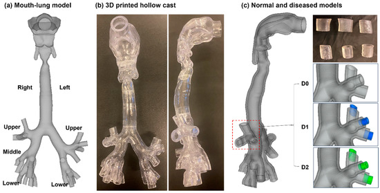
In this study, we present a novel approach to differentiate normal and diseased lungs based on exhaled flows from 3D-printed lung models simulating normal and asthmatic conditions. By leveraging the sequential learning capacity of the Long Short-Term Memory (LSTM) network and the automatic feature extraction of convolutional neural networks (CNN), we evaluated the feasibility of the automatic detection and staging of asthmatic airway constrictions. Two asthmatic lung models (D1, D2) with increasing levels of severity were generated by decreasing the bronchiolar calibers in the right upper lobe of a normal lung (D0). Expiratory flows were recorded in the mid-sagittal plane using a high-speed camera at 1500 fps. In addition to the baseline flow rate (20 L/min) with which the networks were trained and verified, two additional flow rates (15 L/min and 10 L/min) were considered to evaluate the network’s robustness to flow deviations. Distinct flow patterns and vortex dynamics were observed among the three disease states (D0, D1, D2) and across the three flow rates. The AlexNet-LSTM network proved to be robust, maintaining perfect performance in the three-class classification when the flow deviated from the recommendation by 25%, and still performed reasonably (72.8% accuracy) despite a 50% flow deviation. The GoogleNet-LSTM network also showed satisfactory performance (91.5% accuracy) at a 25% flow deviation but exhibited low performance (57.7% accuracy) when the deviation was 50%. Considering the sequential learning effects in this classification task, video classifications only slightly outperformed those using still images (i.e., 3–6%). The occlusion sensitivity analyses showed distinct heat maps specific to the disease state. Full article
Show Figures

Figure 1

---

Figure 3

---

Figure 4

---

Figure 5

---

Figure 6

---

Figure 7

---

Figure 8

---

Figure 9

---

Figure 10

14 pages, 857 KiB  
Review
Primary Resistance to EGFR Tyrosine Kinase Inhibitors (TKIs): Contexts and Comparisons in EGFR-Mutated Lung Cancer
by Keigo Kobayashi
J. Respir. 2023, 3(4), 223-236; https://doi.org/10.3390/jor3040021 - 13 Dec 2023
Viewed by 1245
Abstract
The discovery of the efficacy of tyrosine kinase inhibitors (TKIs) in epidermal growth factor receptor (EGFR)-mutated non-small-cell lung cancer (NSCLC) patients has revolutionized lung cancer therapy. Although almost all responders acquire drug resistance within a few years, many studies have revealed [...] Read more.
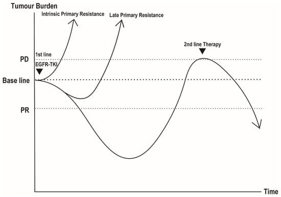
The discovery of the efficacy of tyrosine kinase inhibitors (TKIs) in epidermal growth factor receptor (EGFR)-mutated non-small-cell lung cancer (NSCLC) patients has revolutionized lung cancer therapy. Although almost all responders acquire drug resistance within a few years, many studies have revealed several acquired-resistant mechanisms and developed therapeutic strategies countervailing them, most notably against the EGFR T790M gatekeeper mutation. However, little progress has been made in terms of elucidating the mechanisms of primary resistance. Primary resistance may be defined into two types of resistance, clinically representing patients that do not respond (non-responders) to EGFR-TKIs. The first group consists of approximately 10% of patients that are insensitive to EGFR-TKIs from the outset (intrinsic primary resistance), and 20–30% of the second group consists of patients that seem to clinically benefit at first, but experience early relapse within six months (late primary resistance). In this review, we first provide an overview of drug-induced lung cancer dynamics. We then delve into the mechanisms of primary resistance, with a primary focus on two specific subtypes of resistance. We suggest that “intrinsic primary resistance” is characterized by pre-existing somatic and genomic changes and cell of origins, while “late primary resistance” is correlated with the drug-tolerant persister state. Developing therapeutic strategies to overcome primary resistance is crucial to prolonging the duration of EGFR-TKI therapy. Ultimately, this will allow for an enhanced understanding of lung cancer’s evolutional process, leading to the reversal of acquired resistance and the complete eradication of lung cancer. Full article
Show Figures

Figure 1

EGFR-mutated non-small-cell lung cancer undergoing EGFR-TKI treatment. The post-treatment dynamics of EGFR-mutated non-small cell lung cancer patients receiving EGFR-TKI therapy can be broadly categorized into three groups. The first group is referred to as “intrinsic primary resistance”, where patients show resistance to EGFR-TKI even before treatment initiation. The second group is known as “late primary resistance”, in which patients initially respond to EGFR-TKI therapy for a short period but quickly develop resistance. The third group represents the typical treatment course, with patients undergoing approximately a one-year period of EGFR-TKI exposure before acquiring resistance.

Full article ">
Back to TopTop